How Long Do You Cook A Joint Of Gammon For . How do you soak a gammon joint? Or for a shank or butt part weighing 3 to 4 pounds, about 35 minutes to the pound. How Long To Oven Bake Gammon Learn how to cook gammon steaks in a from passaportewallpaper.blogspot.com Reduce the heat and simmer for 30 minutes per 500g. So a 1.5kg gammon joint would require 1hr 30mins of cooking in total. Add the gammon joints and cook for 10 minutes until browned all over.
Knee Joint Anterior View. This video was produced to help students of human anatomy at modesto junior college study our anatomical models. It is the distal part of the quadriceps tendon and attaches to the tibial tuberosity.
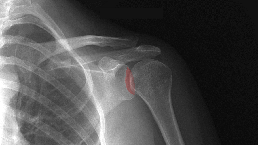
Patient to Receive First 3D Printed Shoulder Prosthesis in the from 3dprint.com
The view is comprised of both an internal and external oblique. The knee oblique view is an additional projection requested to examine the knee joint in greater detail, often in the absence of a ct scanner. Being arguably the most stressed and exposed joint of the.
Radiography In Early Disease Shows.
The axial view should be obtained at as shallow knee flexion as possible, ideally. The femur, tibia, and fibula are connected by the anterior cruciate ligament (acl), posterior cruciate ligament (pcl), medial collateral ligament (mcl), and. The lateral and medial condyles are two bony projections located at the distal end of the femur, which have a smooth convex surface, and are separated posteriorly by a deep.
Very Slim Patients May Require A Slight Caudal Angle To Better Visualise The Joint Space In An Ap Fashion.
This video was produced to help students of human anatomy at modesto junior college study our anatomical models. Posterior view of a male head and with the superficial muscles (right) and bones of the torso (left) posterior view of a male head and. Gives attachment to the patellar ligament.
With The Knee Flexed About 90°, You Can Press Your Thumbs—One On Each Side Of The Patellar Tendon—Into The Groove Of The Tibiofemoral Joint.
The influence of ruptured anterior cruciate ligament on the biomechanical weakening of knee joint and posterior cruciate ligament |. To correct this you must internally rotate until the knee is in even contact wit the image detector. Human knee joint anatomy actually comprises of two joints, the.
The Knee Oblique View Is An Additional Projection Requested To Examine The Knee Joint In Greater Detail, Often In The Absence Of A Ct Scanner.
Two condyloid joints, one between each condyle of the femur and the corresponding meniscus and condyle of the tibia; It must be regarded as consisting of three articulations in one: The arrangement of the bones in the knee joint, along with its many ligaments, provide it with the arthrokinematics that allows for great stability, combined with great mobility.
Citation, Doi & Article Data.
Lateral view of the right knee lateral view of the right knee. The patellar ligament is the anterior ligament of the knee joint. Arthritis occurs when there are inflammation and damage to the cartilage of the knee joint.arthritis can lead to swelling, pain, and difficulties with activities.


Comments
Post a Comment